Magnetic resonance imaging (MRI) is a technique commonly used to explore the brains of sheep. Until now, it had only been performed under general anesthesia, to ensure the animal’s immobility. Anesthesia, however, leads to stress and other negative side-effects, in addition to jeopardizing the study of brain activity.
A research team from INRAE has developed a training protocol adapted to sheep in order to carry out MRI acquisitions in animals while they are awake, without the need for restraint. To do this, researchers drew on previous work with dogs, which until now had been the only animal species capable of carrying out this type of protocol.
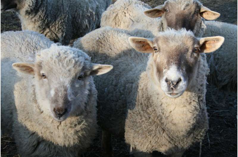
In the nursery of the Animal Physiology Facility experimental research unit (UEPAO), located at the INRAE Val de Loire center in Nouzilly, researchers began a familiarization phase as soon as the lambs were born. The objective was to identify which animals were most receptive to being stroked or to having foam objects placed near their heads.
The paper is published in the journal Behavior Research Methods.
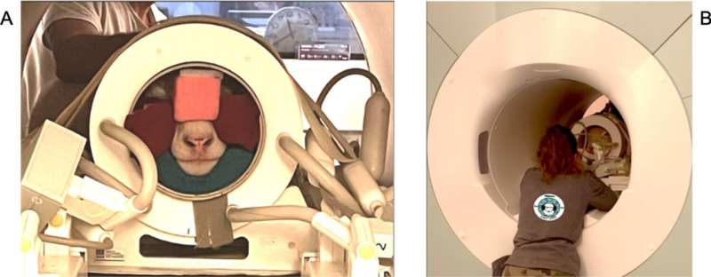
After choosing 10 lambs, an initial training phase took place at the Nouzilly sheep farm. The research team trained the animals to climb a ramp to reach a mock MRI scanner and then lie down. The lambs were also taught to place their heads in a mock MRI coil.
Once they arrived at the real MRI room, the sheep were able to reproduce the same behavior very easily, but had some difficulty remaining perfectly still. It took a few weeks for the animals to get used to the vibrations of the machine and stop moving for a few minutes. Ultimately, the MRI images of their brains were comparable to those obtained from anesthetized sheep, a goal that was initially achieved in six out of the ten sheep trained at the time of writing, and has since been achieved in nine sheep. The protocol lasted nine months, from the birth of the lambs to the first MRI acquisitions.

The success of this protocol is already opening up new avenues for research into animal neuroimaging (e.g., fMRI)—since it makes it possible to study brain function in awake animals. A study looking into the activation of certain brain regions in relation to hearing is currently underway, and is the subject of a Ph.D. thesis that relies on this training protocol.
This example of voluntary cooperation between trainer and sheep illustrates the animal’s ability to learn, and underlines the importance of human-animal relationships in the development of innovative methods. The study also opens up new possibilities for training other animals to carry out awake MRI scans. Such training methods could have numerous other applications, in areas such as shearing or medical training—when the animal learns to collaborate during veterinary care.
More information:
Camille Pluchot et al, Sheep (Ovis aries) training protocol for voluntary awake and unrestrained structural brain MRI acquisitions, Behavior Research Methods (2024). DOI: 10.3758/s13428-024-02449-6
Provided by
INRAE – National Research Institute for Agriculture, Food and Environment
Citation:
Researchers train sheep to complete awake MRI imaging (2024, July 1)
retrieved 1 July 2024
from https://phys.org/news/2024-07-sheep-mri-imaging.html
This document is subject to copyright. Apart from any fair dealing for the purpose of private study or research, no
part may be reproduced without the written permission. The content is provided for information purposes only.


